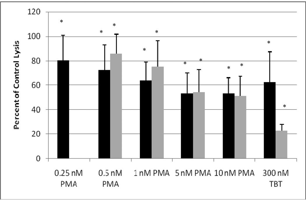
Figure 1.
Effects of exposures to PMA on the ability of NK cells to lyse tumor cells. NK cells were exposed to 0.25–10 nM PMA or 300 nM TBT for 1 h (black bars) or exposed to 0.5–10 nM PMA or 300 nM TBT for 1 h followed by a 24 h period in PMA-free media prior to assaying for NK lytic function (gray bars): To combine results from separate experiments (using cells from different donors) the levels of lysis were normalized as the percentage of the lytic function of the control cells in a given experiment. Values are mean±S.D. from four separate experiments using different donors (triplicate determinations for each experiment, n = 12). An asterisk indicates a significant decrease as compared to control (p<0.05).

