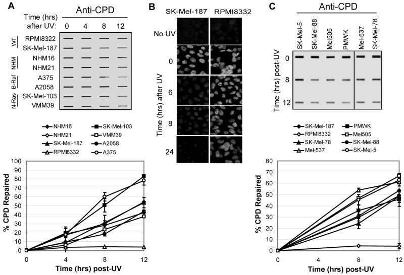
Figure 3. CPD repair in melanocytes and melanoma cell lines.
(A) Cells were exposed to UVC (10 J/m2) and harvested at the indicated time points for immunoslot blot analysis of CPD repair. The top image shows a representative experiment. CPD repair assays were performed three times for each cell line and average CPD repair (and standard deviation) graphed below. (B) Immunofluorescence analyses of CPD repair in SK-Mel-187 and RPMI8332 cells. (C) CPD repair in six additional melanoma cell lines with wild-type N-Ras and B-Raf. The graph (right panel) includes SK-Mel-187 and RPMI cells analyzed in (A).

