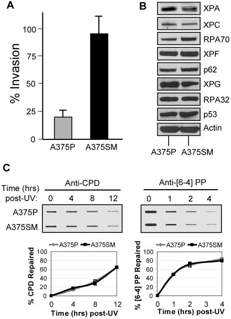
Figure 5. Excision repair in highly metastatic melanoma cells.
(A) Matrigel invasion assay of the highly metastatic cell line (A375SM) compared with its parent cell line (A375P). The results are the averages and standard deviations of three independent experiments. (B) Western blot analyses of the DNA excision repair protein levels of both A375P and A375SM cell lines. (C) Immunoslot blot analysis of CPD and [6-4] PP repair after UV irradiation (10 J/m2) of A375P and A375SM cells. The graphs show the average and standard deviation from two independent experiments.

