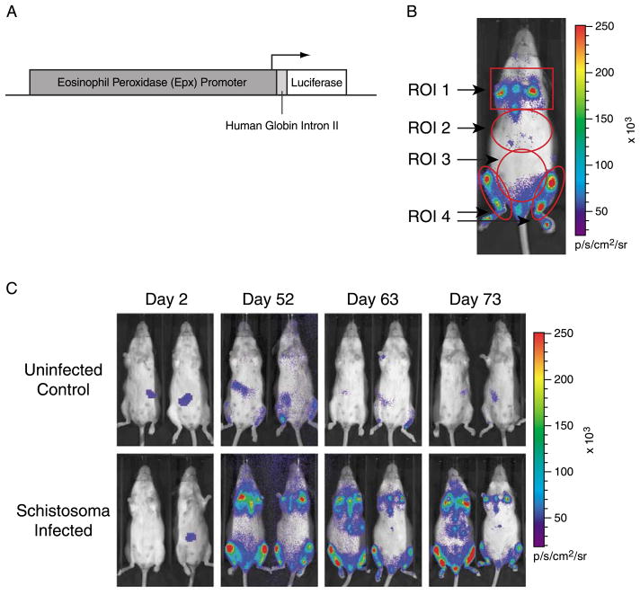
Fig. 1.
In vivo bioluminescence imaging of luciferase reporter gene signal in EPX-luc mice. Luciferin was administered to anesthetised EPX-luc mice and bioluminescence was detected and quantified 10–20 min later using an IVIS® Imaging System 100 Series. (A) schematic of the transgene construct used to generate EPX-luc reporter mice. (B) representative EPX-luc animal, at 70 days p.i. with Schistosoma mansoni, showing the locations of ROI 1 (sternum and forelimb long bones), ROI 2 (liver), ROI 3 (intestine) and ROI 4 (hindlimb long bones). (C) representative uninfected and schistosome-infected EPX-luc animals at various time points p.i. False color overlay represents bioluminescence intensity in photons/s.

