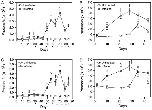
Fig. 5.
In vivo bioluminescence in the long bones and sterna of Schistosoma mansoni-infected EPX-luc mice. In vivo bioluminescence in uninfected and schistosome-infected EPX-luc mice was detected twice weekly over the first 12 weeks of infection as described in Fig. 1. Data from all time points were quantified using LivingImage software and represented graphically. (A) Quantified image data from ROI encompassing the hindlimb long bones (ROI 4) during the 12-week course of the study. (B) Expanded view of hindlimb (ROI 4) data during the first 43 days of the study. (C) Quantified image data from ROI encompassing the forelimbs and sternum (ROI 1) during the 12-week course of the study. (D) Expanded view of forelimb and sternum (ROI 1) data during the first 43 days of the study. ▲: one control and two infected mice euthanised for histology and in vitro tissue luciferase assays. Time points where statistically significant differences in bioluminescence between infected and uninfected animals were detected by Mann–Whitney sign rank test are denoted by *P≤0.05; †P≤0.01; ‡ P≤0.001; and §P≤0.0001. Mean readings±SEM are shown.

