Figure 3.
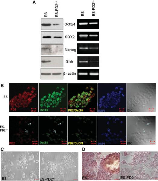
Expression of self-renewal markers and morphological variation of Paf1/PD2 knockout and wild type ES cells. (A): Expression of Oct3/4, SOX2, Nanog, and Shh proteins and mRNA in Paf1/PD2 knockout ESCs and wild-type (WT) ESCs by Western blot and reverse-transcription polymerase chain reaction analysis. (B): Confocal analysis to show Paf1/PD2 and Oct3/4 to localization in WT ESCs and Paf1/PD2 knockout ESCs. The arrows indicate the decreased expression of PD2 and Oct3/4 in Paf1/PD2 knockout ESCs. (C): Phase-contrast images of control and Paf1/PD2 knockout ESCs (scale bar = 0.8 mm). (D): Alkaline phosphatase analysis of WT ESCs and Paf1/PD2 knockout ESCs (scale bar = 0.8 mm). Abbreviations: DAPI, 4′,6-diamidino-2-phenylindole; DIC, differential interference contrast; PD2+/−, PD2 heterozygous knockout.

