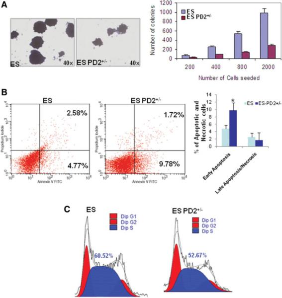
Figure 4.
Loss of Paf1/PD2 abolishes ESC functions. (A): Qualitative and quantitative analysis for short-term colony formation in WT and Paf1/PD2 knockout ESCs. (B): Annexin V and propidium iodide staining analysis by flow cytometry to identify apoptosis in WT ESCs and Paf1/PD2 knockout ESCs. The statistical analysis showed significant (*, p = .05) variation in early apoptosis but not in late apoptosis or necrosis in Paf1/PD2 knockout ESCs. (C): Cell cycle analysis of WT ESCs and Paf1/PD2 knockout ESCs by flow cytometry using propidium iodide. Abbreviations: FITC, fluorescein isothiocyanate; PD2+/−, PD2 heterozygous knockout.

