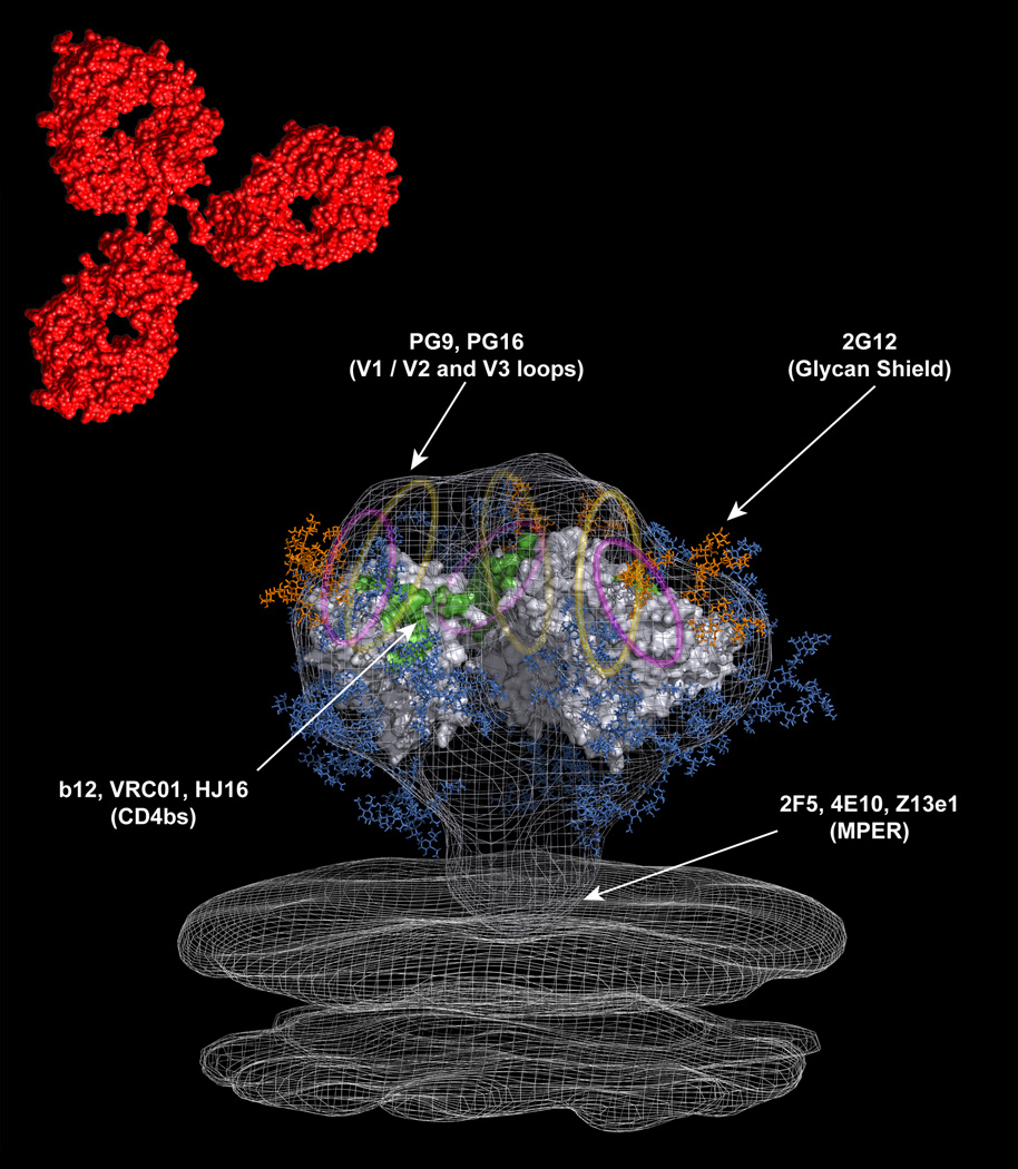
Figure 2. Modeling the epitopes recognized by bNAbs onto the HIV-1 trimer.
The above model is adapted from a recent cryo-electron tomographic structure of the HIV-1 trimer [28••,86]. The crystal structure of the b12-bound monomeric gp120 core has been fitted into the density map [39]. The V1/V2 and V3 loops, which are not resolved in the structure, are represented as yellow and magenta ovals, respectively. The red structure located above the trimer is representative of a human IgG1 molecule. The approximate locations of the epitopes targeted by the existing bNAbs are indicated with arrows. Carbohydrate chains are shown in blue, and the oligomannose cluster targeted by mAb 2G12 is shown in orange.

