
Figure 4.
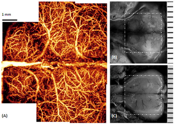
The entire cerebro-vascular flow over the cortex of an adult mouse with the skull intact was imaged with OMAG in vivo. (A) is the projection view of blood perfusion that reveals the detailed blood perfusion network over the cortex at capillary level resolution. (B) Photography of the skull with the skin folded aside, taken right after the OMAG imaging where viewing the vasculature through the skull is not possible. (C) Photography of blood vessels over the cortex, after the skull and the meninges of the same mouse were carefully removed. The superficial major blood vessels are in agreement with those in OMAG image in (A). The area marked with the dashed white box represents 7.5×7.5 mm2.