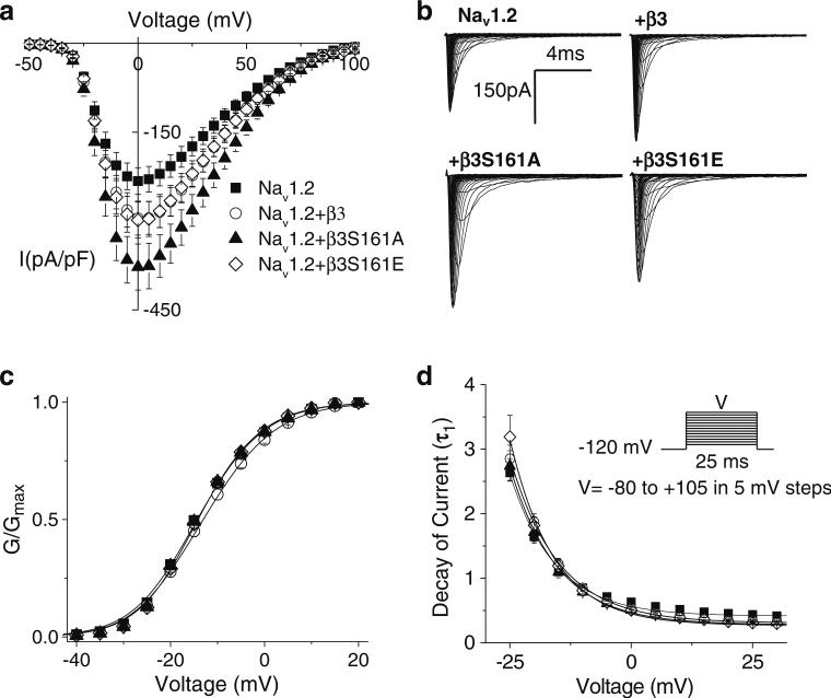
Fig. 1.
β3 co-expression increases current density without modulation of activation parameters. a Current density, assessed as the peak of the current voltage relationship for each cell divided by cell capacitance, was amplified by β-subunit co-expression. Currents were elicited by depolarizing steps of 25 ms from a holding potential of –120 mV to test pulses in the range of –80 to +105 mV in 5-mV increments. Co-expression of β3(β3–306.1±29.3 pA/pF, n=57) or β3S161E (–303.4±30.4 pA/pF, n=39) increased the median current density as compared to non-transfected cells (Nav1.2=–235.3±22.5 pA/pF, n=48). Co-expression of β3S161A resulted in a significantly greater median current density than non-transfected cells (–389.9±34.5 pA/pF, n=44, Dunn's test p<0.05). b Representative traces of whole-cell Na currents evoked in nontransfected HEK293 cells stably expressing Nav1.2 and cells co-transfected with WTβ3, β3S161A, and β3S161E. c Conductance plot shows no effects on activation gating parameters with any of the β-subunit constructs. Smooth lines correspond to the least squares fit when average conductance data were fit with a single Boltzmann equation. d Decay of macroscopic currents was fit to double exponential function and was not significantly different across the three expression systems. Data shown are means ± SEM

