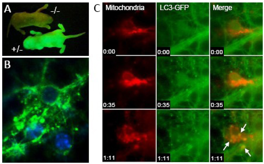
Fig. 2.
Dynamic tracking of autophagy in primary cortical neurons in vitro. A. Discrimination of GFP-LC3+/− mice using fluorescent macroscopy. B. Identification of GFP-LC3-enriched vesicles suggestive of autophagosomes in primary cortical neurons from GFP-LC3+/− mice after 24 hours of nutrient deprivation using fluorescent microscopy. C. Dynamic tracking of mitophagy in primary cortical neurons from GFP-LC3+/− mice using time-lapsed microscopy. Autophagosomes (green) fusing with mitochondria (red; MitoTracker Red, Invitrogen, Carlsbad, CA) are highlighted by arrows. Time stamp is hours:minutes.

