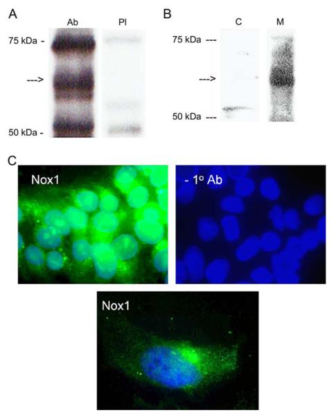
Figure 2.
(A) Western blot of Nox1 (arrow) in BeWo cell lysate employing anti-Nox1 serum (Ab) or pre-immune serum (PI). (B) Western blot of Nox1 (arrow) in BeWo cell protein fractions. C, cytosolic protein; M, membrane protein. (C) Fluorescent immunohistochemistry of Nox1 expression in BeWo cells detected by FITC-conjugated anti-rabbit IgG. Cell nucleuses were stained with DAPI.

