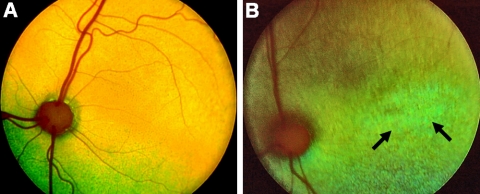
Figure 2.
Fundus photographs of a 12-week-old normal Abyssinian cat (A) and an affected mixed breed (Rdy) cat (B). The generalized grayish discoloration of the fundus in the affected cat is most notable in the area centralis, as is the generalized vascular attenuation. Arrows: marked changes in the area centralis of the affected cat.

