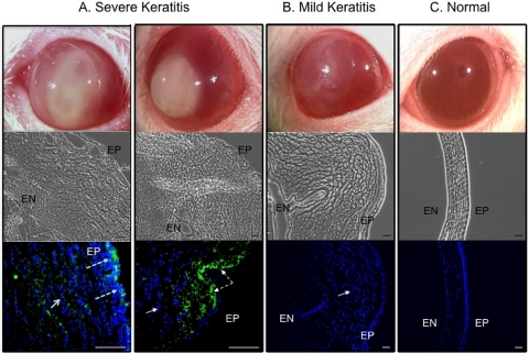
Figure 4.
Representative photographs and cross-sectional phase-contrast and fluorescence micrographs of contact lens–induced P. aeruginosa keratitis in the rat model of extended wear. (A) Severely diseased eyes from two rats with total scores of 10 or above at day 5 (showing a lens still in place) and day 6 after inoculation, respectively, (B) a mildly diseased eye with a total score below 10 at day 7 after inoculation (showing the lens in place), and (C) an uninoculated normal eye (contralateral control). Corneal and infiltrating cell nuclei were stained with DAPI (blue). All diseased eyes showed large numbers of infiltrating cells within the cornea. Severely infected corneas had the worst structural disruption and showed GFP-expressing P. aeruginosa derived from original inoculum (green). EP, corneal epithelium; EN, endothelium. Scale bar, 50 μm. Images were taken from the experiments shown in Figure 2 (high-inoculum group).

