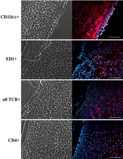
Figure 5.
Immunostaining of infiltrating cells (red) in cross-sections of a rat cornea at day 3 after inoculation with P. aeruginosa in the lens-wearing model. Keratitis was moderate. Right: infiltrating cells labeled with mouse monoclonal antibodies against rat CD11b/c (CR3 complement receptor, i.e., phagocytes), ED-1 antigen (monocytes, macrophages), αβ T-cell receptor (mature and immature αβ T cells), or CD4. Cell nuclei were counterstained with DAPI (blue). Left: phase-contrast images of the corresponding tissue sections. Scale bar, 50 μm. Images were taken from the experiments shown in Figure 2 (low-inoculum group).

