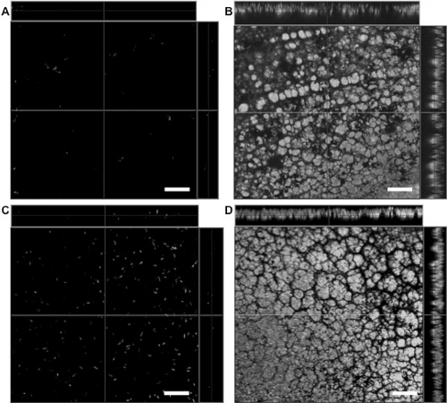
Figure 6.
Laser scanning confocal microscopy of P. aeruginosa-inoculated contact lenses before and after in vivo wear in the rat contact lens model. The anterior surface of a worn lens (A) and posterior surface of the same lens (B) after 4 days of wear in the rat eye (low-inoculum group). Classical biofilm architecture of GFP-expressing P. aeruginosa strain PAO1 (derived from the original inoculum) was found on the posterior lens surface (B). Bacteria were found on the posterior lens surface at the time of fitting (C) (∼105 cfu). A classical biofilm of GFP-expressing P. aeruginosa strain PAO1 was also found on the posterior surface of lenses worn for 9 days (D) (high inoculum). Scale bar, 20 μm.

