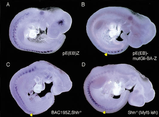
Figure 5.
Analysis of reporter gene transcription. In situ hybridization of nlacZ transcripts was performed on 9.5-dpc pE(EB)Z (A), pE(EB)mutGli-BA-Z (B), and BAC195Z, Shh-/- embryos (C). In situ hybridization of Myf5 transcripts was performed on 9.5-dpc Shh-/- embryos (D). In wild-type pE(EB)Z embryos, nlacZ transcripts are detected in all epaxial somites, although less strongly at rostral levels (A). In contrast, in wild-type pE(EB)mutGli-BA-Z, nlacZ transcripts are seen predominantly in the youngest somites (arrowhead in B). In BAC195Z, Shh-/- and Shh-/- embryos, nlacZ and Myf5 transcripts, respectively, are detected in ventral dermomyotomes and in poorly developed myotomes along the rostro-caudal axis. Although corresponding β-galactosidase activity is detected by Xgal staining, both transcripts are visualized in a fuzzy pattern in the epaxial somites in the mutants (arrowheads).

