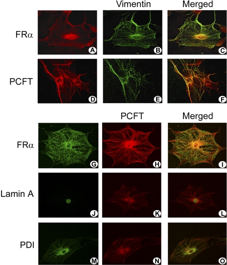
Figure 4.
Laser-scanning confocal microscopic immunolocalization of FRα and PCFT in primary Müller cells. Müller cells that were freshly isolated from mouse retina were grown on coverslips and subjected to immunofluorescent detection of FRα (red, A) and vimentin (green, B) or PCFT (red, D) and vimentin (green, E). Areas of colocalization of the folate transport proteins with vimentin are shown (merged images, orange-red, C, F). Immunodetection of FRα (green, G), PCFT (red, H), and coimmunolocalization (merged image, orange, I). Detection of the nuclear marker lamin A (green, J), PCFT (red, K), and coimmunolocalization (merged image, orange, L) and the ER marker PDI (green, M), PCFT (red, N), and coimmunolocalization (merged image, orange, O).

