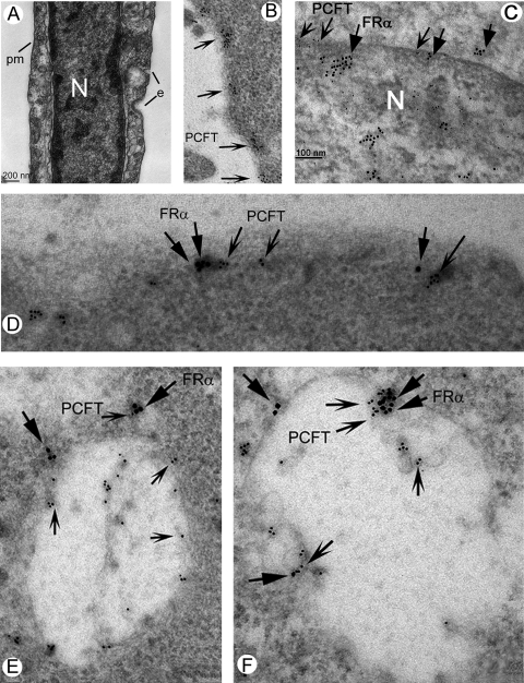
Figure 5.
Electron microscopic immunolocalization of FRα and PCFT in Müller cells. Müller cells were fixed, and postembedding electron microscopy immunolocalization was used to detect PCFT (10-nm gold particle, indented arrows) and FRα (18-nm gold particle, flat arrows). (A) Electron microscopic photomicrograph (lower magnification) of a Müller cell. The nucleus (N) is prominent in the cell. e, Endosomes forming along the plasma membrane (pm). (B) PCFT immunolabeling on the plasma membrane. (C) PCFT and FRα immunolabeling along the nuclear membrane. (D) PCFT and FRα immunolabeling along the plasma membrane. (E, F) Immunodetection of PCFT and FRα in endosomes; note several areas in which the two proteins are in proximity.

