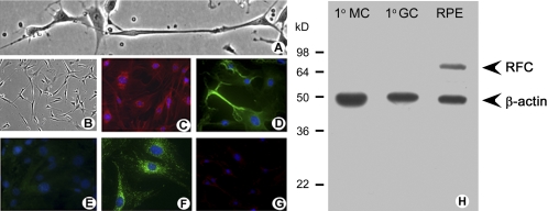
Figure 8.
Immunocytochemical and Western blot analysis of FRα and RFC in freshly isolated retinal Müller cells. (A) Phase-contrast microscopic analysis of primary Müller cells in culture. (B) Lower magnification of the cells shown in (A). Cells were processed for immunofluorescence detection of (C) CRALBP (red), (D) vimentin (green), (E) NF-L (green, negligible in these cells), (F) FRα (green), and (G) RFC (green, negligible in these cells). In all immunofluorescence experiments, DAPI was used to stain the nuclei of the cells (blue). (H) Primary Müller cells, primary ganglion cells, and RPE/eyecup protein lysates were prepared from mouse and used to perform Western blot analysis. RFC (∼65 kDa) was detected in RPE, which served as a positive control. RFC was not expressed in the primary Müller cells (1°MC) or primary ganglion cells (1°GC). β-Actin (∼45 kDa) served as the loading control.

