
Figure 4.
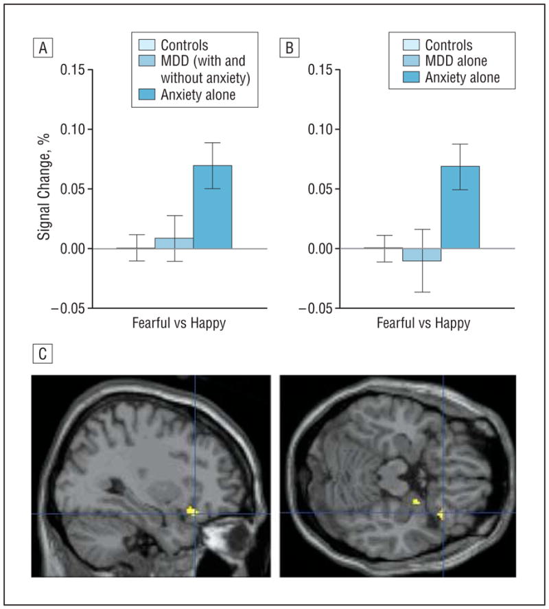
Orbitofrontal cortex (OFC) activation in the “fearful-passive vs happy-passive” contrast in patients with major depressive disorder (MDD) and anxiety. A and B, Right OFC activation to fearful faces during passive viewing of fearful vs happy faces (fearful-passive vs happy-passive contrast) (error bars reflect standard errors) showing significantly enhanced activation among patients with anxiety compared with those with MDD and healthy controls. C, The fearful-passive vs happy-passive contrast evidences significantly greater right lateral OFC activation in patients with anxiety than in those with MDD (with and without comorbid anxiety) (Montreal Neurological Institute coordinates: 32, 24, −18, P=.005, small-volume corrected; no suprathreshold voxels emerge for the anxiety vs MDD-alone comparison). Highlighted areas indicate regions where the differences in blood oxygen level–dependent activation between groups were significant (for display purposes, uncorrected threshold was set at P=.0005).