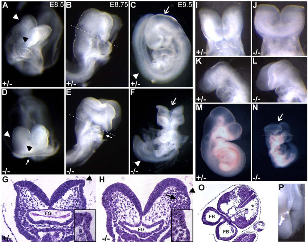
Fig. 3.
Morphological defects of the Ovol2 mutant embryos. Morphological comparison of control (A–C) and mutant (D–F) B6 × 129 embryos at the indicated stages. (A, D) Morphology at E8.5. Note the absence of a heart in this mutant embryo (arrow). (B, E) Slightly later in development, the defects in the presumptive brain and heart became more apparent. (C, F) At E9.5, the cranial neural tube region of the mutant embryos was still open (arrow). Note the twisted appearance of the trunk in (F) (arrowhead). (G–H) Histological sections through the hindbrain region of E8.75 embryos as indicated in (B) and (E). The arrowheads demarcate the neuroectoderm/surface ectoderm junction. The arrow in (H) indicates cells with neural crest cell morphology in closer proximity to the neuroectoderm/surface ectoderm border in the mutant. Inset panels in (G) and (H) represent higher magnification images of the neuroectoderm/surface ectoderm transition. FG, foregut. (I–P) Morphological analysis of E8.5 (I–L) and E9.5 (M–P) embryos in the CD1-enriched genetic background. Ventral (I–J) and lateral (K–L) views of control and mutant embryos at E8.5 revealed enlarged, round neural folds in the mutant. Note the open cranial neural tube (arrow) in the mutant at E9.5 (N). (O) Section through the cranial region of a mutant embryo (plane of cleavage is indicated in N). FB, forebrain. (P) Dorsal view of the trunk region of an “escaper” that displayed spina bifida.

