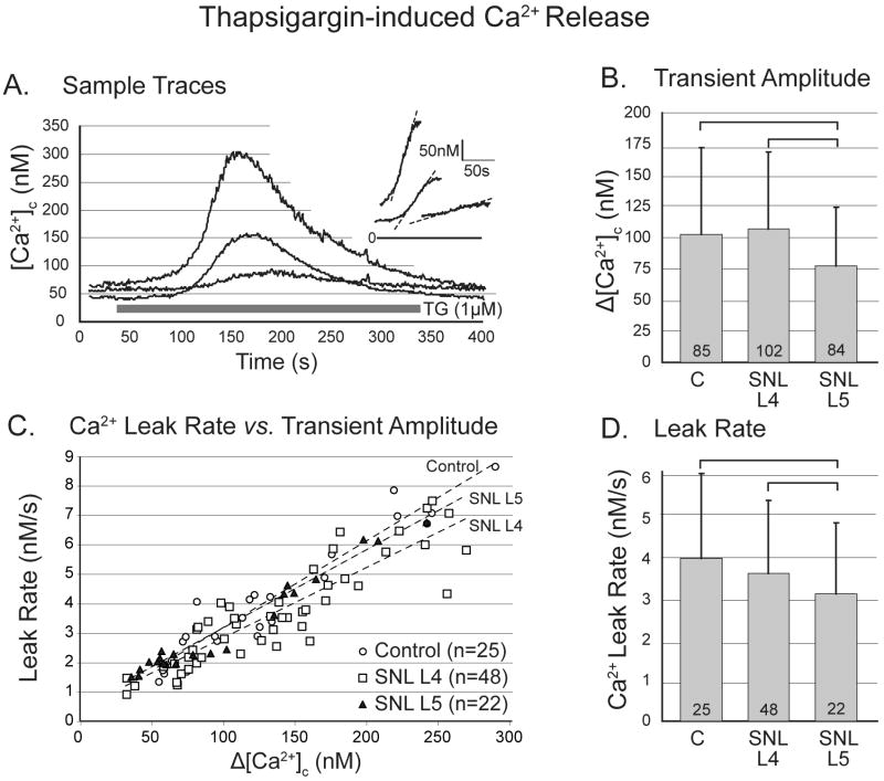
Figure 3.
Response of cytoplasmic Ca2+ concentration ([Ca2+]c) in sensory neurons to application of thapsigargin (TG). A. Sample traces from control (C) neurons demonstrate variable amplitude of response but a linear rising limb of the transient (inset). B. Spinal nerve ligation (SNL) depresses the response in axotomized neurons of the fifth lumbar (L5) dorsal root ganglion. (One-way ANOVA with Bonferroni post-hoc testing.) C. The rate of Ca2+ leak during TG application, measured as the slope of the rising limb, is linearly dependent on the transient amplitude for all injury groups. D. Leak rate is depressed in L5 neurons. For panels C and D, an ANCOVA model with Bonferroni post-hoc testing was employed. For panels B and D, numbers in the bars indicate n for number of neurons, brackets above bars connect groups that are significantly different (P<0.05), and error bars show standard deviation.

