
Figure 2.
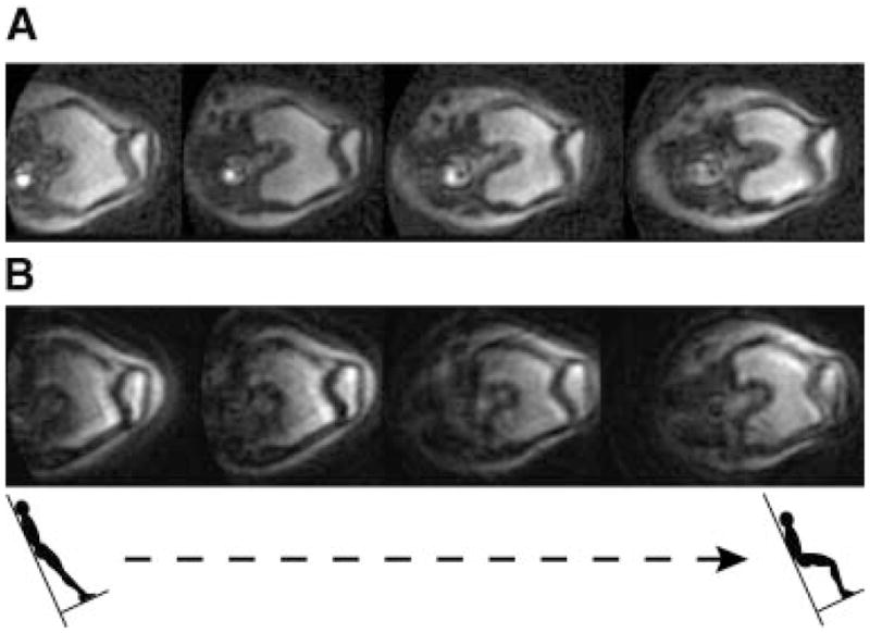
(A) Real-time MR images of the patellofemoral (PF) joint of a pain-free control subject during upright, weight-bearing knee extension. (B) Real-time MR images of the PF joint of a subject with pain during upright, weight-bearing knee extension. Notice the lateral position and rotation of the patella relative to the femur as the pain subject nears full extension. These are oblique-axial views through the knee corresponding to four knee flexion angles between 0 and 60°.