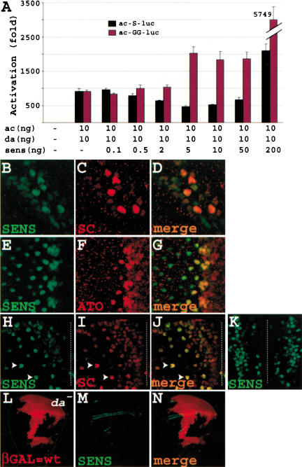
Figure 6.
Sens acts as a dose-dependent transcriptional repressor and activator. (A) Transcription assays with higher proneural-to-Sens ratio show that Sens can act as a repressor. In addition, mutation of the Sens-binding site abolishes the repressive ability of Sens and causes synergy at levels that repress the wild-type enhancer. (B-K) Sens is not only expressed in SOPs but also in surrounding cells in the wing margin (B-D) and the eye imaginal disc (E-G) and pupal microchaetae field (H-K). Note that Sens is expressed at low levels in the nuclei of many cells in the wing margin of a Canton S third instar larva (B), and in 6-7 cells in each preommatidial cluster in the eye disc of a Canton S third instar larva (E). (H-J) Part of the hemi-notum of an 8-10-hour-old sensE2/+ pupa, which essentially has a wild-type bristle pattern. The future midline is depicted by the broken line. Note the field of cells closer to the midline that express low levels of Sens and Sc. Arrowheads show two examples of presumptive SOPs, which have up-regulated Sens and Sc. Note that the cells surrounding the SOPs show minimal Sens and Sc expression at this stage. (K) Part of the notum of a 7-9-hour-old Canton S pupa containing the medial aspects of both developing hemi-nota. Note columns of Sens-expressing cells on both sides of the midline. (L-N) Sens expression in the wing margin does not depend on da. Large mitotic clones for a null da allele are evident by the lack of βGAL staining (L). Note that in both the large clone covering the anterior margin and the small clone touching the posterior margin Sens expression is intact (N). Also note that in precursors of the ventral and dorsal radii, there seems to be a mutant cell at the border of the clone with Sens expression.

