
Figure 5.
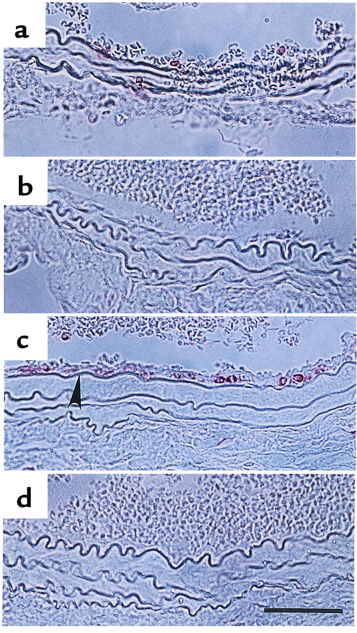
CD45 staining of paraffin sections from the mouse carotid artery after ligation. (a and b) 3 days after ligation; (c and d) 7 days after ligation. (a and c) Mdk+/+ mice; (b and d) Mdk–/– mice. Arrowheads indicate CD45-positive cells. Bar, 50 μm.