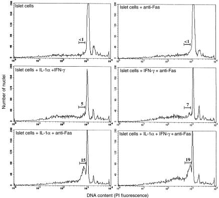
Figure 3.
Susceptibility of cytokine-treated islet cells to Fas-induced apoptosis. Islets from RAG-2–/– NOD mice were cultured for 16 hours as above in the presence or absence of IL-1α, IFN-γ, IL-1α + IFN-γ, and anti-murine Fas mAb. Islets were then dispersed into single cells, fixed in ethanol, stained with propidium iodide (PI), and analyzed by flow cytometry to determine the presence of apoptotic nuclei. The numbers above the bars indicate the percentage of cells with hypodiploid DNA content (early apoptotic cells).

