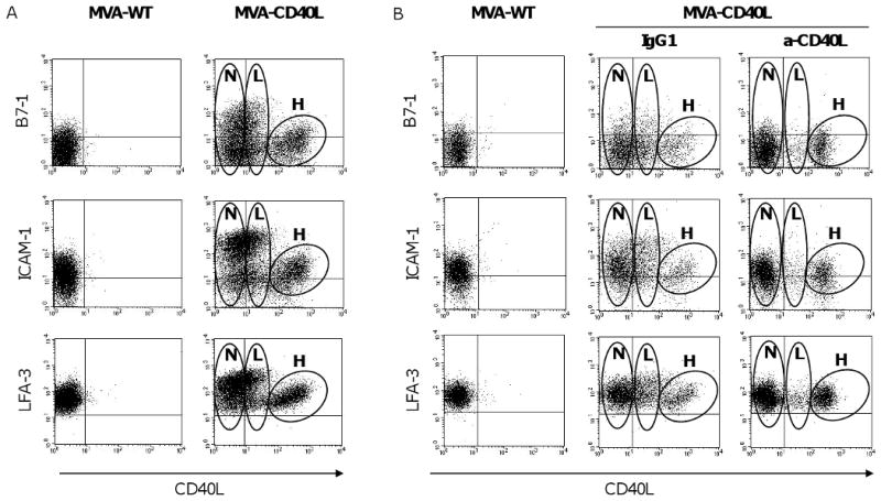
Figure 2. Upregulation of costimulatory molecules by CLL cells following MVA-CD40L infection.

(A) CLL cells were infected with MVA-WT or MVA-CD40L. Following 24 hours of infection, CLL cells were analyzed by flow cytometry for expression of B7-1, ICAM-1, and LFA-3 by CD40L-expressing and non-expressing populations. The plots shown were gated on CD19+ B cells. After infection with MVA-CD40L, 3 populations of CLL cells that expressed negative, low, or high levels of CD40L were observed; these are designated as “negative” (N), “low MFI” (L), and “high MFI” (H) populations on the plots shown. (B) CLL cells were treated with blocking antibody (anti-CD40L or isotype-control mouse IgG1) before and during infection with MVA-CD40L.
