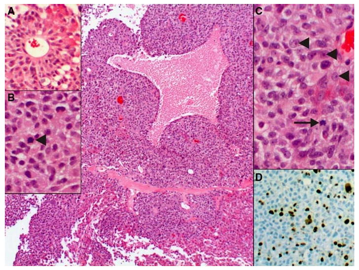
Fig. 1.

Pituitary tumor, arranged in sheets of pleomorphic epithelial cells embedded in a delicate capillary network. Areas of coagulative necrosis were identified, most likely due to infarction secondary to the large size of the tumor. a In areas, pseudorosette-formation around larger caliber vessels was present. b The cells have a moderate to abundant amount of granular eosinophilic cytoplasm, round to oval nuclei with many mitotic figures (arrowhead). c The neoplastic cells have distinct, eosinophilic nucleoli (arrowheads) and hyperchromatic chromatin. Mitotic figures were prominent (arrow). d The high proliferation rate as indicated by the elevated mitotic count was corroborated by a high Ki67 (mib-1) labeling index, reaching up to 31% of the cells
