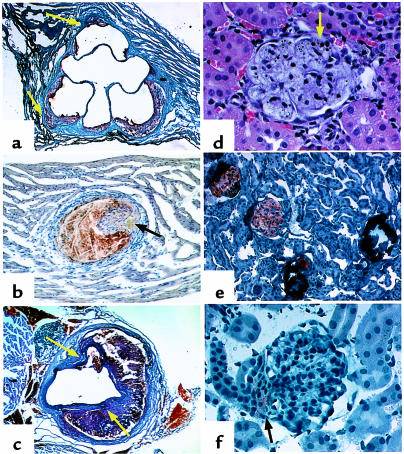
Figure 3.
Representative histological sections from 4-month-old nnee animals. (a) Proximal aorta section showing atherosclerotic lesions. Sudan IVB with hematoxylin counterstain; ×40. Arrows indicate microaneurysms. (b) A plaque in a small vessel in myocardium. Sudan IVB with hematoxylin counterstain; ×200 (initial magnification). Arrow indicates lumen. (c) Aneurysms in an abdominal aortic section. Sudan IVB with hematoxylin counterstain; ×40 (original magnification). Arrows indicate sites of dissection. (d) Foam cells in glomerulus. Hematoxylin and eosin; ×165. Arrow indicates small area of calcification. (e) Four glomeruli demonstrating the transition from heavy lipid deposition (which appears orange) to dystrophic calcification (which appears dark blue). Sudan IVB with hematoxylin counterstain; ×82.5. (f) Glomerulus of enalapril-treated nnee mouse. Lipid deposition is light (arrow) and is confined to extraglomerular mesangial cells. Sudan IVB and hematoxylin; ×165. This pattern of lipid staining is similar to that seen in NNee mice.

