
Figure 2.
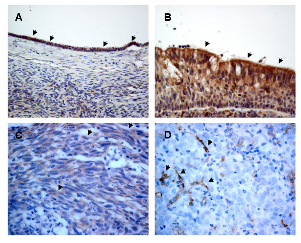
Immunohistochemical analysis of DAB2 expression in NPC. (A) Surface epithelium of ovary served as positive control (original magnification × 400). (B) Normal nasopharyngeal epithelium (× 400). Arrows indicate the positive epithelium. (C) Cytoplasmic staining in a NPC biopsy (× 200). Arrows indicate positive NPC cells. (D) Negative staining in NPC cells. The dendritic cells (arrows) in the stroma served as internal positive controls (× 200).