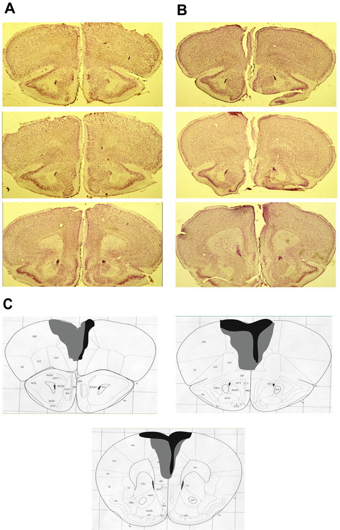
Figure 3.
Histological verification of the extent of medial prefrontal cortex damage. A. Representative sections from three coordinates along the anterior-posterior axis of the mouse prefrontal cortex in a sham-operated animal. B. Corresponding sections from an animal given ibotenic acid infusions into the medial prefrontal cortex. C. A diagram shows the extent of the largest (light gray) and smallest (black) lesion across the fourteen animals.

