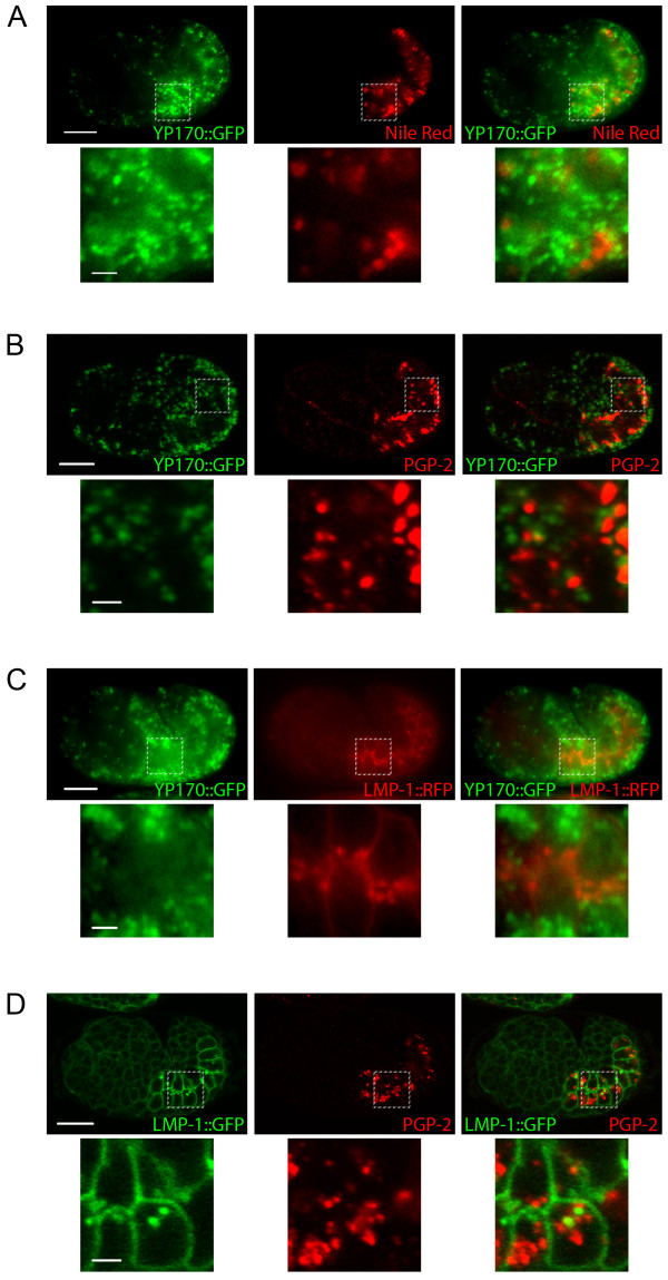
Figure 1.
Late endocytic and lysosome-related organelles in developing intestinal cells of wild type embryos. A) Epifluorescence images of a wild type embryo that expresses YP170::GFP and stained with Nile Red. B) Confocal images of a wild type embryo that expresses YP170::GFP and immunostained to detect PGP-2. C) Epifluorescence images of a wild type embryo that expresses YP170::GFP and LMP-1::TagRFP(S158T). D) Confocal images of a wild type embryo that expresses YP170::GFP and immunostained to detect PGP-2. Bottom panels are magnified images of the regions indicated in the top panels. Scale bars in whole embryo images represent 10 μm; scale bars in magnified images represent 2 μm.

