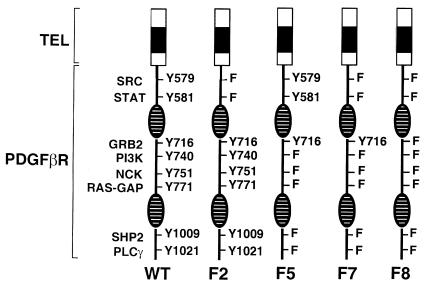
Figure 4.
Tyrosine to phenylalanine signaling mutants of TEL/PDGFβR. The NH2-terminal TEL portion of the fusions, indicated by a shaded box, remains unchanged in the wild-type and mutants. Phosphotyrosines in the PDGFβR portion of the fusion are indicated by “Y” and are numbered according to the wild-type PDGFβR sequence. Mutants were generated with tyrosine to phenylalanine (F) substitutions at specific residues as indicated. The split kinase domain of PDGFβR is indicated by striped ovals.

