Figure 7.
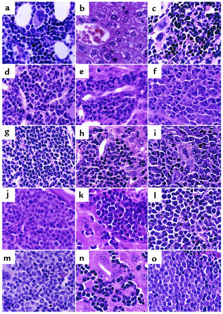
Histopathology of mice transplanted with TEL/PDGFβR and related mutants. High-power (×640) views of H&E-stained bone marrow (a, d, g, j, m), liver (b, e, h, k, n), and spleen (c, f, i, l, o) of transplanted mice. (a–c) Negative control mouse transplanted with the ΔPNT mutant of TEL/PDGFβR demonstrates normal tissues after transplant. (a) Intramedullary adipocytes and a normal mixture of maturing myeloid and erythroid precursors and scattered megakaryocytes are seen in the bone marrow. (b) A central venule surrounded by normal hepatocytes. Splenic red pulp (c) demonstrates a mixture of maturing erythroid and myeloid elements similar to that seen in the marrow. TEL/PDGFβR (d–f) and F5 mutant (g–i) tissues both demonstrate marked expansion of myeloid precursors (identified as cells with folded or ringlike nuclei) within bone marrow and splenic red pulp, as well as marked extramedullary hematopoiesis comprising mainly myeloid precursors within the liver. In contrast, F2 (j–l), F7 (m–o), and F8 (not shown) tissues show a monomorphous population of lymphoblasts that efface the marrow and splenic red pulp and infiltrate liver sinusoids.

