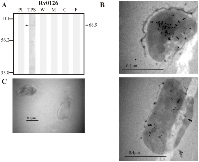
Figure 11. Experimental assessment of the subcellular localization of the negative control protein Rv0126.
(A) Immunoblotting assessment of the presence of Rv0126 in TPS: total protein sonicate, W: cell wall, M: membrane, C: cytosol and F: culture filtrate of M. tuberculosis H37Rv, using specific antisera raised in rabbits. PI: assessment of the pre-immune serum showing no recognition of any mycobacterial protein. Molecular weight marker is shown on the left (P7708S ColorPlus Prestained Protein Marker, New England Biolabs) and the molecular weight observed for Rv0126 is shown to the right. (B) IEM assessment of the presence of Rv0126 in the cytoplasm of intact M. tuberculosis H37Rv bacilli (magnification: 40,000×). Proteins detected by anti-rabbit antibody conjugated to 10-nm colloidal gold particles are indicated by the black arrows. (C) Pre-immune serum showed no recognition of any mycobacterial proteins. The results showed detection of Rv0126 in TPS and the cytoplasm of M. tuberculosis H37Rv.

