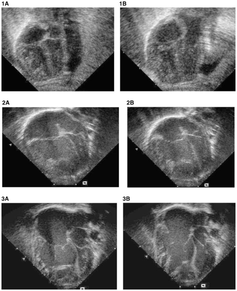
Figure 1–3.

This series of four-chamber two-dimensional echocardiographic images compares the disproportion between right ventricle and left ventricle in congenital diaphragmatic hernia (Fig. 1A, B); total anomalous pulmonary venous drainage (Fig. 2A, B); and cerebral arteriovenous malformation (Fig. 3A, B) in systole (Fig. 1,2,3A) and diastole (Fig. 1,2,3B). The enlarged apex-forming right ventricle compresses the left ventricle. The left ventricle in these conditions differs from true HLHS because the aortic and mitral valves are morphologically normal, and when pulmonary hypertension resolves, the left ventricle assumes a normal shape and volume.
