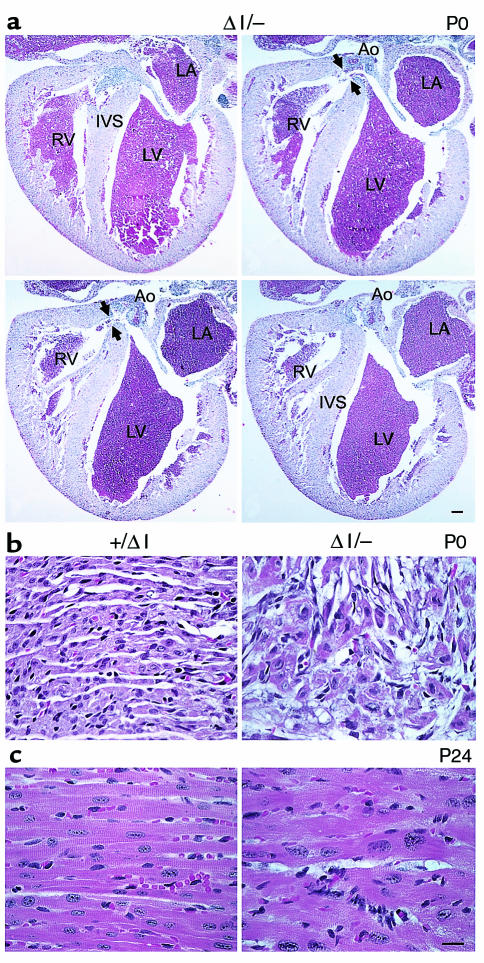
Figure 6.
Histological sections of BΔΙ/B– and B+/BΔΙ mouse hearts. (a) Four selected serial sections, anterior to posterior (clockwise from upper left), demonstrating the defect (arrowheads) in the membranous region of the ventricular septum in a P0 BΔΙ/B– mouse heart. Ao, aorta; IVS, interventricular septum; LA, left atrium; LV, left ventricle; RV, right ventricle. H&E stain. Bar, 200 μm. (b) Normal cardiac histology in a B+/BΔΙ mouse (left) and area of myocyte disarray and hyperchromatic nuclei in a BΔΙ/B– mouse (right). (c) Hypertrophied myocytes from a P24 BΔΙ/B– mouse (right) and myocytes from a B+/BΔΙ littermate (left). H&E stain. Bar, 20 μm.

