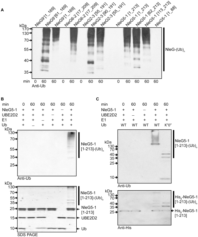
Figure 4. In vitro activity of NleG proteins.
(A) Immunoblot analysis using anti-ubiquitin antibodies of reactions performed in the presence of ATP, ubiquitin, E1, UBE2D2 and full length or C-terminal fragments of His6-NleG isoforms. (B) Immunoblot analysis with anti-ubiquitin antibodies (top panel) and SDS PAGE electrophoresis (bottom panel) of reactions performed with ATP and in the presence or absence of ubiquitin, E1, UBE2D2 E2 and NleG5-1[1–203]. (C) Immunoblot analysis with anti-ubiquitin antibodies (top panel) and anti-His6 antibodies (bottom panel) of reactions performed with E1, wild type ubiquitin (WT) or ubiquitin derivative (K”O”) in the presence or absence of His6-NleG5-1[1–203] and UBE2D2 E2 enzyme. All reactions were incubated at 30°C for the times indicated.

