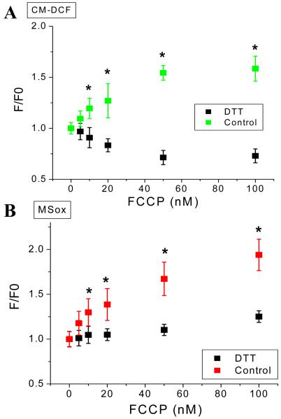
Figure 11. Mild uncoupling can increase or decrease mitochondrial ROS depending on the redox environment in isolated cardiomyocytes from guinea pig heart.
Freshly isolated cardiomyocytes from guinea pig heart were handled, and loaded with the ROS probes CM-H2DCFDA (A) and MitoSOX (B) and imaged with two photon scanning laser fluorescence microscopy as described in Methods and the legend of Figure 8. After baseline imaging of cells in the absence or the presence of 1mM or 2mM dithiothreitol (DTT) perfused with tyrode pH 7.5 containing 1mM Ca2+ and 10mM glucose, the indicated nanomolar concentrations of the protonophore FCCP were added. Identical imaging protocol to that described in Figure 8 was followed in the FCCP dose response. Data for control or DTT pretreated cells were paired, i.e. performed in the same cells at all uncoupler concentrations, all throughout the experiment (n=6 for each FCCP concentration; 2 experiments). * p < 0.05.

