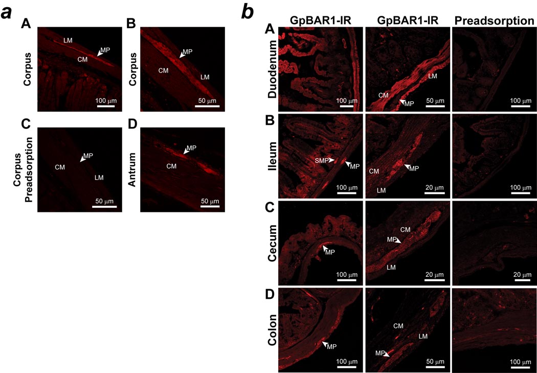
Figure 2. Localization of GpBAR1-IR in sections of mouse stomach and small and large intestine.
a: Representative examples of GpBAR1-IR in the wall of the gastric corpus (A, B, C) and antrum (D). GpBAR1-IR was expressed in the myenteric plexus (MP). Circular muscle, CM; longitudinal muscle, LM. Preadsorption of the GpBAR1 antibody with immunizing peptide abolished GpBAR1-IR (C).
b: Representative examples of GpBAR1-IR in the wall of the duodenum (A), ileum (B), cecum (C) and colon (D) are shown. GpBAR1-IR was expressed in the myenteric plexus (MP) and submucosal plexus (SMP) of the small and large intestine. GpBAR1-IR was more highly expressed in the mucosa, muscularis externa (circular muscle, CM; longitudinal muscle, LM), of the small intestine compared to the large intestine. Preadsorption of the GpBAR1 antibody with immunizing peptide abolished GpBAR1-IR.

