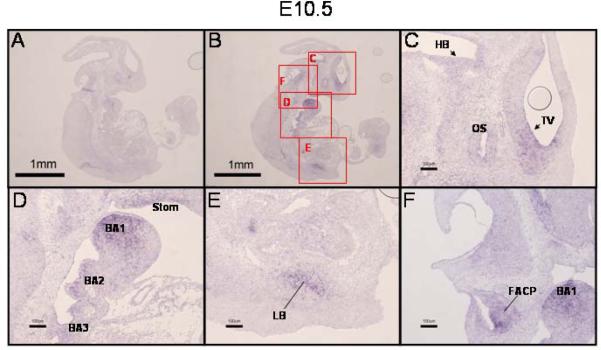
Figure 2. Expression of PRDM16 in the E10.5 mouse embryo.
A,B) In situ hybridization with PRDM16 sense or antisense riboprobe, respectively (low magnification, 20X). Lettered red boxes in panel B indicate areas shown in higher magnification in panels C–F (100X). C) Similar to E9.5, expression of PRDM16 was detected in the neuroepithelium lining telencephalic vesicle (TV, arrowhead). (Note: the circle within the TV in panel C is an artifact (air bubble)). Expression of PRDM16 was also detected in the neuroepithelium of the hindbrain (HB, arrowhead) and in the optic stalk (OS). D) Notable expression of PRDM16 was seen in the first, second and third branchial arches (BA1, BA2, BA3) and in the epithelium of the stomadeal roof (Stom). PRDM16 was expressed in the developing lung bud (LB, panel E) and in the facio-acoustic preganglion complex (FAPC, panel F). Scale bar = 1 mm in panel A and B and 100 μm in panels C–F.

