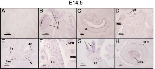
Figure 5. Broad expression of PRDM16 in E14.5 mouse embryos.
In situ hybridization of sagittal sections of an E14.5 mouse embryo with a PRDM16 sense (A) and antisense (B–H) riboprobes. Expression of PRDM16 during this stage of development encompasses multiple tissues including widespread signals in the developing brain (Br, panel B) and olfactory epithelium (OE, panel C). Panel D demonstrates expression of PRDM16 in the trigeminal ganglion (TNG), the vestibulocochlear ganglion (VIII), and submandibular glands (SMG). Expression was also observed in the developing testis (Te), pancreas (Pan), kidney (Ki) and adrenal gland (AG) (panel E). The developing lung (Lu) also expressed PRDM16 as well as the dorsal root ganglia (DRG) and cartilage primordia of the ribs (CPR) (panel F). In the hindlimb, PRDM16 expression was restricted to the perichondrium surrounding the developing tarsal bones (panel G, arrows). Expression was detected in the eye in the pigment layer of the retina (PLR) and in the lens (Lens, panel H). Scale bars: panels A and B = 1 mm; C, F, and H = 100 μm; and D, E, and G = 400 μm.

