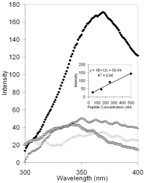
Figure 2.

Intrinsic Fluorescence of the DNA-PNA-peptide-PEG Conjugate. The purified DP3 conjugates and conjugates prepared identically but in the absence of one component were analyzed via intrinsic fluorescence spectroscopy to detect tryptophan residues present in the linker peptide (excitation 280 nm; emission 350 nm). The DP3 conjugate (black diamonds) exhibited significant fluorescence at 350 nm, indicative of the presence of the linker peptide. In contrast, samples prepared in the absence of DNA (grey squares), PNA (grey “X”s), or peptide (grey triangles) had minimal fluorescence at the same wavelength. Inset: the fluorescence emission peak intensities of various linker peptide solutions were analyzed (excitation 280 nm; emission 350 nm).
