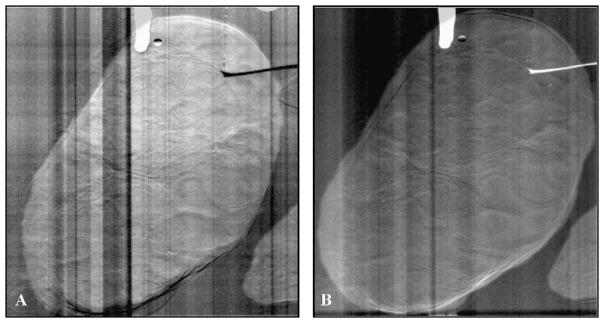
Fig. 13.
Diffraction enhanced images (DEI) of a bovine ovary produced with X-rays on beamline ×15A of the National Synchrotron Light Source, Brookhaven National Laboratory, Upton, NY. Three antral follicles approximately 4 mm in diameter are indicated on the left, and a corpus luteum is outlined on the right. The images were acquired on opposite sides of the diffraction rocking curve, resulting in opposing contrast. Artifact was produced by a paper clip (at top) used to suspend the ovary in a plexiglass water bath (vertical striations).

