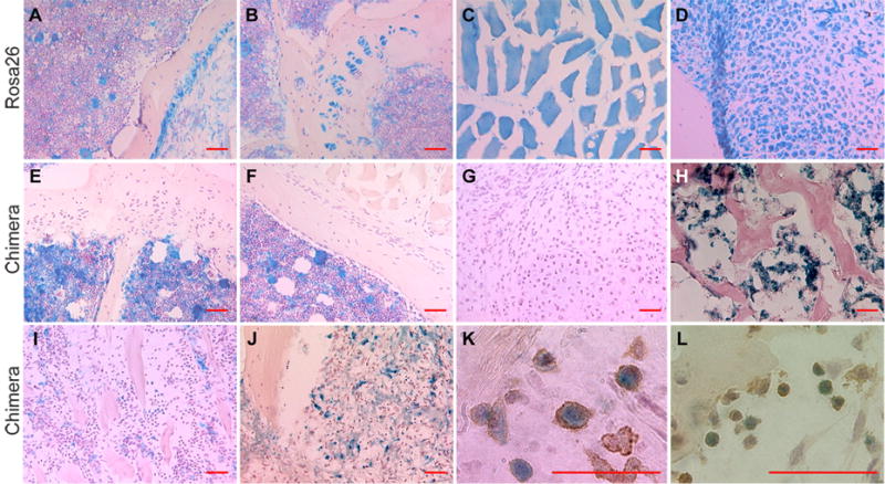
Figure 2. Distribution of donor bone marrow derived cells in the chimeras.
(A) Cells in bone marrow, (B) periosteum, growth plate, (C) muscle fibers, and (D) chondrocytes in the fracture callus were stained blue by X-gal staining in ROSA26 mice. (E) In chimeric mice, donor cells were not observed in growth plate, (F) cortical bone, periosteum, muscle, and (G) cartilage and (H) bone in the callus. (I) 7 days after fracture donor cells (blue) were present at the fracture site and (J) in granulation tissue. (K) Donor cells were stained with F4/80 or (L) MCA771G. (Scale bar = 50μm)

