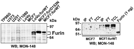
Figure 7. Analysis of cellular furin.
Left panel, Western blotting of total cell furin (15 µg total protein that corresponded to 5-7×10⁴ cells depending on a cell type). Right panel, Western blotting of cell surface-associated furin. MCF-7 and MCF-7:furWT cells (15×106) were cell surface biotinylated using membrane-impermeable biotin. Biotin-labeled furin was immunoprecipitated from the total cell lysate (TCL) using streptavidin-agarose beads. The beads were washed in 50 mM Tris-HCl, pH 7.4, supplemented with 50 mm N-octyl-β-d-glucopyranoside, 150 mM NaCl, 1 mm CaCl2, 1 mm MgCl2, a proteinase inhibitor cocktail set III, and 1 mm PMSF (FT, flow through fraction). The immunocaptured proteins (IP, immunoprecipitated protein fraction) were eluted using 1% SDS. The fractions were analyzed by Western blotting with the furin MON-148 antibody followed by donkey anti-mouse IgG-conjugated with horseradish peroxidase and a SuperSignal West Dura Extended Duration Substrate kit. The gels were overexposed to demonstrate the presence of cell-surface furin. Right lane, purified furin (1 ng). WB, Western blotting.

