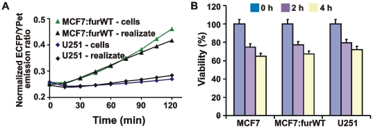
Figure 11. Furin activity was released by the cells.
A, the time course of the biosensor cleavage by cell realizate and by adherent MCF-7:furWT and U251 cells. Cells (5×10⁴) were incubated for 2 h in 100 mM Hepes, pH 7.5, containing 150 mM NaCl, 1 mM CaCl2, 1 mM MgCl2 and 1% ITS. The cells were then separated by centrifugation and the supernatant (realizate) was co-incubated with the biosensor for 0–120 min. Alternatively, adherent cells (5×10⁴) were directly co-incubated with the biosensor. B, ATP-Lite cell viability assay. Prior to the assay, MCF-7, MCF-7:furWT and U251 cells were incubated for 2–4 h at 37°C in 100 mM Hepes, pH 7.5, containing 150 mM NaCl, 1 mM CaCl2, 1 mM MgCl2 and 1% ITS. The level of induced apoptosis was then determined using an ATP-Lite kit.

