
Figure 4.
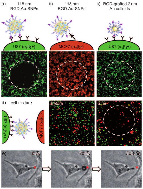
a–c) Fluorescence micrographs of U87 cells (αvβ3 +, labeled green) treated with 118 nm RGD-Au-SNPs (a), MCF7 cells (αvβ3-, labeled red) treated with 118 nm RGD-Au-SNPs (b), and U87 cells treated with RGD-grafted 2 nm Au colloids (c) after irradiation with a pulsed laser (6 ns, 120 mJcm−2). A mask was employed to confine the laser beam to a circular region with a diameter of 1 mm (as indicated by the white dashed circles). d) Fluorescence micrographs of a 1:1 mixture of U87 and MCF7 cells. After treatment with RGD-Au-SNPs and subsequent medium exchange, the cell mixture was irradiated with a pulsed laser. In the irradiated region after culture for 2 h, U87 cells (green) were depleted, whereas MCF7 cells (red) were left alive on the substrate. e) Time-resolved images of an αvβ3-positive U87 cell with a 118 nm RGD-Au-SNP attached to it. Upon irradiation with the 6 ns pulsed laser (120 mJcm−2), fast contraction of the cellular protrusion was observed as the result of the localized mechanical destruction caused by the formation of microbubbles.