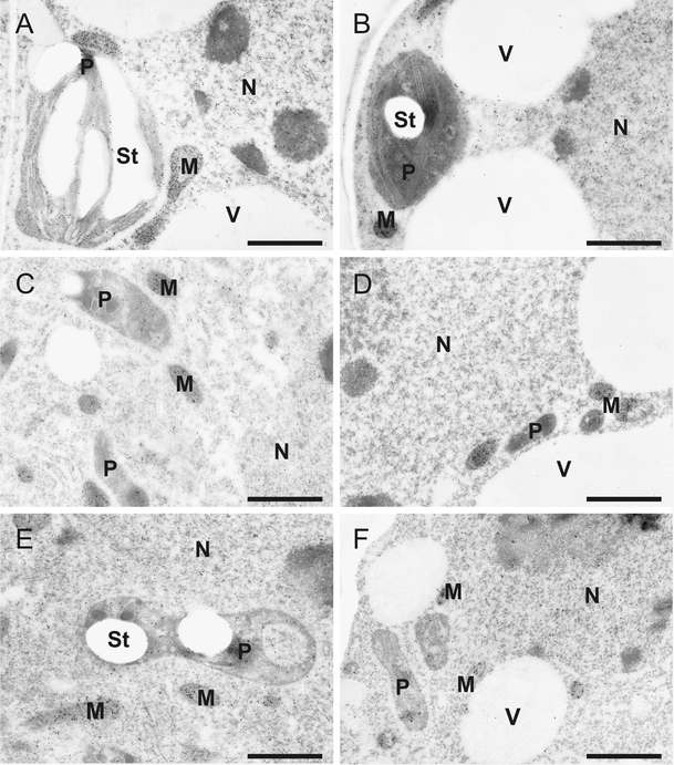
Fig. 1.

Immunogold-labeling of glutathione. Transmission electron micrographs show gold particles bound to glutathione in mesophyll (a, b), long- (c, d) and short-stalked (e, f) glandular trichome cells from leaves of Cucurbita pepo (L.). Lower gold particle density can be found in cells of plants treated with 50 µm cadmium (b, d, f) in comparison to the control (a, c, e). CW cell walls, M mitochondria, N nuclei, P plastids, St starch, V vacuoles. Sections were post-stained with uranyl acetate for 15 s. Bars 1 µm
